

Skin Care
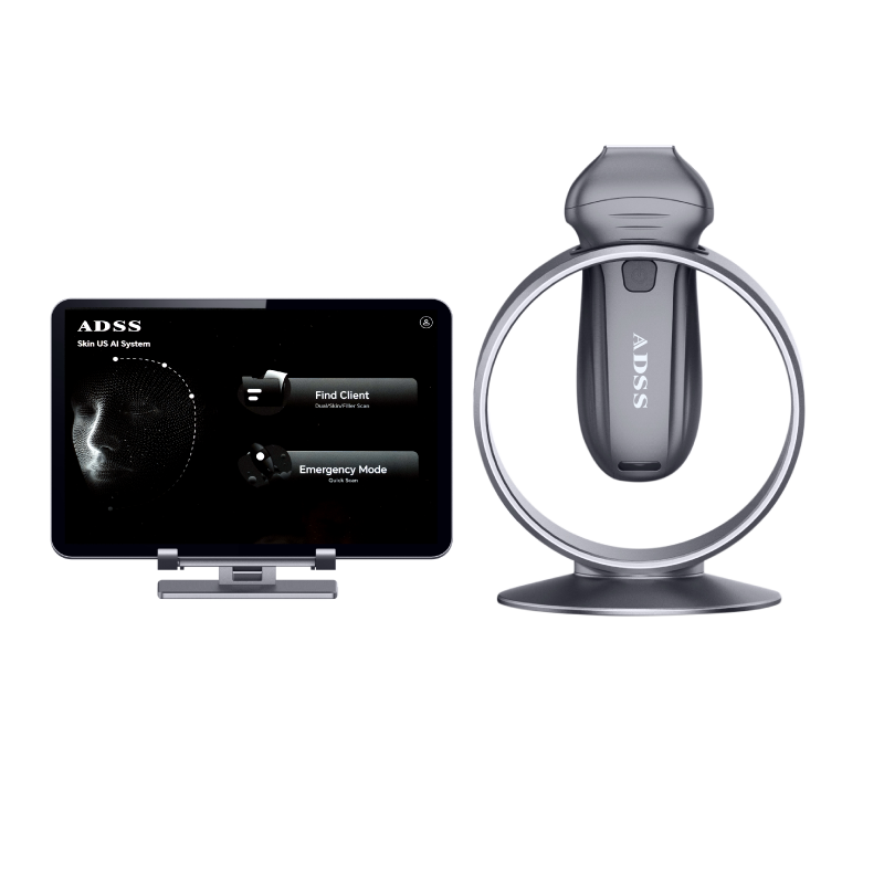
AI Handheld Ultrasound
Full-Layer Skin Imaging Powered by AI.
Product Overview
Product Overview

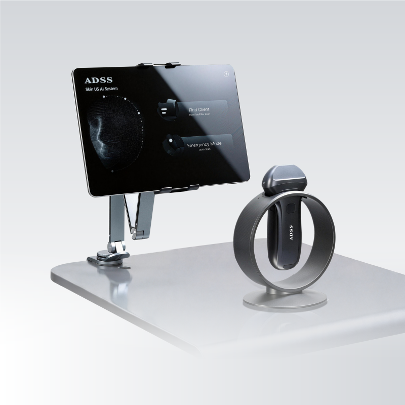
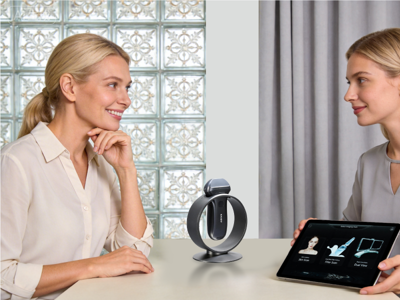
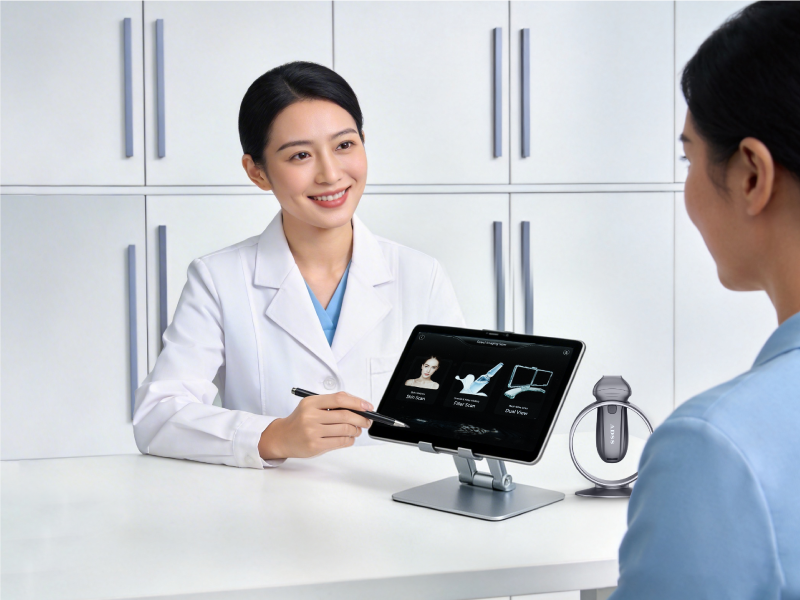
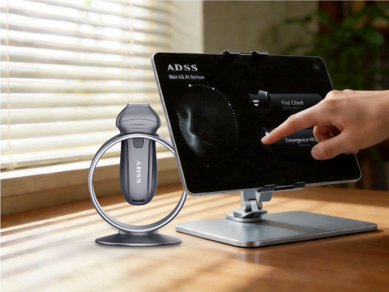
AI Handheld Ultrasound is a compact, professional-grade imaging system designed for aesthetic and dermatological applications. Unlike surface-level skin analyzers, it provides tomographic ultrasound imaging to visualize tissue structures, identify filler distribution, and assess treatment depth in real time.

By combining high-frequency ultrasound imaging with AI-assisted segmentation and recognition, it transforms complex ultrasound data into structured clinical insights, supporting accurate diagnosis, treatment planning, and follow-up comparison.

How It Works

Step 1 — High-Frequency Ultrasound Imaging
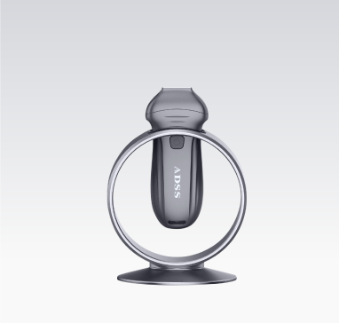
The handheld probe emits 13.2MHz ultrasound waves to penetrate through epidermis, dermis, subcutaneous tissue, and reach the SMAS layer, generating high-resolution acoustic imaging of facial structures. 


Step 2 — AI Real-Time Recognition & Segmentation
The AI engine analyzes ultrasound images in real time, supporting tissue layer segmentation, filler recognition, abnormality screening, and quantitative evaluation. 

Step 3 — Structured Data Output
The system converts raw ultrasound imaging into structured clinical information for evidence-based assessment, including imaging parameters, layer metrics, filler mapping, and longitudinal comparison. 

Step 4 — Multi-Modal Integration (3D Skin Data Compatibility)
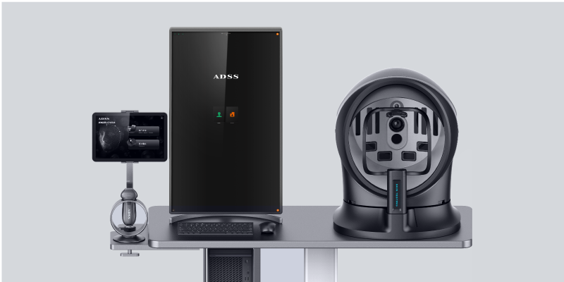
Under online mode, scan data can be synchronized and shared with the 3D skin analysis platform under the same account, enabling cross-platform visualization, comparison, and report generation.


Key Advantages
13.2
MHz
13.2MHz

High-frequency imaging designed for clear visualization from epidermis to SMAS layer.

21
Points
21Points

Standardized facial mapping coverage for full-face scanning without blind spots.

3
Modes
3Modes

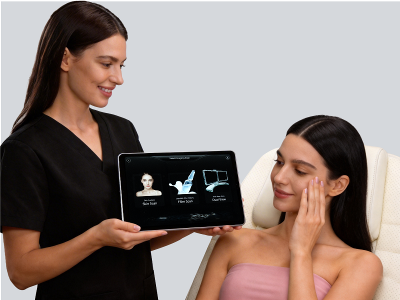
Skin Scan / Filler Scan / Dual View for different clinical needs.

7
Fillers
7Fillers

Supports recognition of 7 common filler materials (HA, Collagen, PLLA, PCL, CaHA, PMMA, PVA).

5
Seconds
5Seconds

Dynamic video capture enables fast clinical imaging and real-time observation.

Real-Time Ultrasound Visualization for Aesthetic Precision.
Full-Layer Visualization to SMAS

Designed with 13.2MHz frequency to achieve clear imaging from epidermis to SMAS layer, allowing professionals to evaluate skin thickness, structure, and deep tissue conditions with confidence. 

AI-Powered Imaging Interpretation

Backed by an AI database of 30,000+ clinical cases, the system performs fast recognition and analysis, reducing dependence on manual interpretation and improving diagnostic consistency. 

Filler Recognition & Injection Safety Support

Supports identification of 7 common filler types, assisting in filler distribution mapping, complication evaluation, and injection depth verification. 

Real-Time Dual View for Dynamic Procedures

Dual-screen real-time imaging supports guidance during injection and treatment, helping practitioners observe needle angle and filler placement dynamically.

Radiation-Free, Non-Invasive Monitoring

Ultrasound-based scanning ensures repeatable use for long-term monitoring without radiation exposure.

Fast Adoption with Minimal Learning Curve

Intuitive UI design supports quick training and efficient clinical workflow, with automated reporting and one-click comparison functions.

Technical Highlights
01
3 Scanning Modes

Skin Scan Mode
Designed for full-layer skin structure analysis.
Visualizes epidermis, dermis, subcutaneous tissue, and SMAS to support skin condition evaluation.

Filler Scan Mode
Designed for filler history analysis and material recognition.
Supports identification of filler distribution, metabolic changes, and abnormality screening.

Dual View Mode
Real-time dual display for dynamic observation.
Ideal for injection guidance and live monitoring during procedures.

02
21 Facial Mapping Points

The system covers 21 standardized facial mapping points.Each point supports structured imaging capture, enabling full-face evaluation with consistent data collection for comparison and reporting.

03
Supported Filler Identification

Recognizable Filler Types
·Hyaluronic Acid (HA)
·Collagen
·PLLA
·PCL
·CaHA
·PMMA
·PVA

The system supports filler recognition and imaging evidence collection for safer aesthetic planning and injection evaluation.

04
AI Database & Accuracy

Powered by an AI database of 30,000+ cases, the system continuously improves recognition performance and adapts to different skin types.
The algorithm accuracy reaches over 96%, supporting more consistent clinical interpretation and reducing diagnostic variability.

05
Imaging Parameter Control

The device supports professional ultrasound parameter adjustment for optimized imaging:
·GN (Gain) – controls brightness
·BDR (Dynamic Range) – controls contrast and grayscale resolution
·D (Depth) – sets scanning depth
·FOC (Focus) – defines focal position
·ENH (Enhancement) – improves edge sharpness
·HAM (Harmonic Imaging) – reduces artifacts and improves SNR
·CPD (Color Power Doppler) – visualizes blood flow signals in real time

06
Custom Acoustic Coupling Pad

The system uses a dedicated acoustic coupling pad designed for consistent ultrasound transmission and stable image quality.

·One client, one pad use
·Enhanced ultrasound clarity and acoustic gain
·No repeated wiping or cleaning required
·More consistent imaging performance compared to traditional gel usage

07
Wireless Workflow & Data Storage

Wireless Scanning
The system uses wireless transmission for real-time ultrasound video streaming and scanning control.

Online Mode
By logging into the online account, ultrasound data can be synchronized with the 3D skin analysis platform under the same clinic account.

Offline / Local Mode
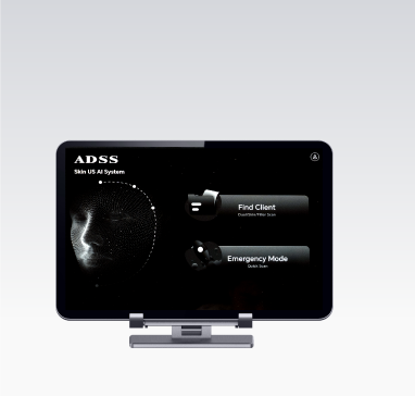
Ideal for exhibitions or clinics without 3D skin analyzers. Data can be stored locally on the tablet. With 256GB storage, it supports up to approximately 2,000 patient scan records, which can later be uploaded manually.

01

3 Scanning Modes

Skin Scan Mode
Designed for full-layer skin structure analysis.
Visualizes epidermis, dermis, subcutaneous tissue, and SMAS to support skin condition evaluation.

Filler Scan Mode
Designed for filler history analysis and material recognition.
Supports identification of filler distribution, metabolic changes, and abnormality screening.

Dual View Mode
Real-time dual display for dynamic observation.
Ideal for injection guidance and live monitoring during procedures.

02

21 Facial Mapping Points

The system covers 21 standardized facial mapping points.Each point supports structured imaging capture, enabling full-face evaluation with consistent data collection for comparison and reporting.

03

Supported Filler Identification

Recognizable Filler Types
·Hyaluronic Acid (HA)
·Collagen
·PLLA
·PCL
·CaHA
·PMMA
·PVA

The system supports filler recognition and imaging evidence collection for safer aesthetic planning and injection evaluation.

04

AI Database & Accuracy

Powered by an AI database of 30,000+ cases, the system continuously improves recognition performance and adapts to different skin types.
The algorithm accuracy reaches over 96%, supporting more consistent clinical interpretation and reducing diagnostic variability.

05

Imaging Parameter Control

The device supports professional ultrasound parameter adjustment for optimized imaging:
·GN (Gain) – controls brightness
·BDR (Dynamic Range) – controls contrast and grayscale resolution
·D (Depth) – sets scanning depth
·FOC (Focus) – defines focal position
·ENH (Enhancement) – improves edge sharpness
·HAM (Harmonic Imaging) – reduces artifacts and improves SNR
·CPD (Color Power Doppler) – visualizes blood flow signals in real time

06

Custom Acoustic Coupling Pad

The system uses a dedicated acoustic coupling pad designed for consistent ultrasound transmission and stable image quality.

·One client, one pad use
·Enhanced ultrasound clarity and acoustic gain
·No repeated wiping or cleaning required
·More consistent imaging performance compared to traditional gel usage

07

Wireless Workflow & Data Storage

Wireless Scanning
The system uses wireless transmission for real-time ultrasound video streaming and scanning control.

Online Mode
By logging into the online account, ultrasound data can be synchronized with the 3D skin analysis platform under the same clinic account.

Offline / Local Mode
Ideal for exhibitions or clinics without 3D skin analyzers. Data can be stored locally on the tablet. With 256GB storage, it supports up to approximately 2,000 patient scan records, which can later be uploaded manually.

New Design

Designed for Professional Aesthetic Imaging Workflows

Functions

Easy generation of structured reports for clinical records


Real-time monitoring to support precise aesthetic procedures


Rapid full-face skin assessment for quick clinical evaluation


Long-term patient data tracking for comparison and outcome monitoring

Treatment Areas

Forehead

Brow area
 

Tear trough / under-eye area
 

Cheeks

Apple muscle zone
 

Nasal bridge

Nasolabial folds

Perioral area

Jawline

Chin

Forehead

Brow area
 

Tear trough / under-eye area
 

Cheeks

Apple muscle zone
 

Nasal bridge

Nasolabial folds

Perioral area

Jawline

Chin

Application Scenarios
Dermatological diagnosis support
Full-layer skin structure analysis
Filler metabolism tracking
Injection and filler evaluation
Facial concern analysis
Post-treatment monitoring and comparison

Dermatological diagnosis support

Full-layer skin structure analysis

Filler metabolism tracking

Injection and filler evaluation

Facial concern analysis

Post-treatment monitoring and comparison

Discover more about ADSS AI Handheld Ultrasound and what it could do for you
Download Brochure
Related Products
Business Value

Evidence-Based Treatment Planning
Provides objective imaging evidence for treatment depth assessment and tissue evaluation.

Safer Injection & Filler Management
Supports filler distribution mapping and needle angle observation in real time, helping reduce misjudgment risks.
 

Longitudinal Monitoring & Before-After Comparison
Repeatable ultrasound scanning enables treatment effect tracking, filler metabolism observation, and structured follow-up.

Business Cooperation
Global Distribution Opportunities with Full Marketing & Technical Support

OEM/ODM services available for private labeling and customization

Regional exclusive distribution rights available

Comprehensive product training, user manuals, operation videos, and sales scripts provided

Marketing toolkit including branded visuals, clinical case videos, and social content

Dedicated technical support team for real-time assistance

OEM/ODM services available for private labeling and customization

Regional exclusive distribution rights available

Comprehensive product training, user manuals, operation videos, and sales scripts provided

Marketing toolkit including branded visuals, clinical case videos, and social content

Dedicated technical support team for real-time assistance

Become a Strategic Global Partner of ADSS
Whether you're a distributor, agent, or brand owner, ADSS offers tailored solutions to scale your business.
Explore Cooperation Models
Download

I consent to receiving communications from ADSS and to the storage and processing of my personal data on the ADSS website. Privacy Policy